


Arthritis is a chronic degenerative disease that affects all parts of the joint: cartilage, articular membrane, ligaments, capsule, periarticular bone and periarticular muscles and ligaments.
According to European doctors, arthrosis accounts for almost 70% of all rheumatological diseases. People aged 40-60 years are most susceptible to joint arthrosis. This is facilitated by both lack of movement and prolonged loads, poor nutrition and, of course, injuries.
What is a joint?
Normally, human joints consist of 2 or more connecting bones. All working surfaces of the joint have a protective coating and are constantly lubricated with synovial fluid for optimal gliding. The joint cavity itself is hermetically sealed by the articular capsule.
In our body, there are many joints that are "responsible" for certain types of movement, can experience various loads and have different safety margins.
The amount of movement in a joint depends on the structure of the joint, the ligamentous apparatus that limits and strengthens the joint, and the various muscles attached to the bone by tendons.
Causes of joint arthrosis
Normal joint function is possible with continuous self-renewal of cartilage tissue. At a young age, the rate of death of outdated joint cells is equal to the rate of birth of new cells. Over the years, the process of cell renewal slows down, and cartilage tissue begins to thin. The production of synovial fluid is also reduced. As a result, the articular cartilage begins to thin and deteriorate, which leads to arthrosis.
In addition, there are other causes of joint arthrosis:
- increased physical activity. Arthritis of the joints is a frequent accompaniment of weight gain. As a result of excessive load, microtraumas are formed in the joints. Athletes experience joint damage due to increased load on "unheated" joints;
- joint injuries;
- congenital or acquired deformities of the musculoskeletal system (rickets, kyphosis, scoliosis, improper fusion of bones after injury with the appearance of limb deformation: O and X-shaped leg deformation).
Stage of arthrosis
Depending on the level of cartilage tissue destruction, different stages or degrees of arthrosis can be distinguished.
Stages and symptoms of arthrosis
- Stage 1 arthrosis is characterized by periodic pain in the joints, especially with increased physical activity. After resting, the pain usually goes away. The range of motion in the joint is not limited, the muscle strength in the injured limb does not change. X-rays can show signs of minimal joint damage.
- Arthrosis of the 2nd degree is indicated by painful sensations not only with intense physical stress, but also with small loads. Even when resting, joint pain may not subside. This stage is characterized by stiffness in movement and limited mobility in the joints. This eventually leads to muscle atrophy. X-rays may show joint deformation, joint space reduction, and the appearance of bone growth near this gap.
- Arthrosis of the 3rd degree - any movement causes a person to experience great pain. Joint pain is present even at rest. Therefore, a person tries to move as little as possible so that the pain is minimal. In some cases, movement requires the use of crutches or a gurney. Sometimes bone fusion occurs - ankylosis (as in ankylosing spondylitis).
With deformed arthrosis, irreversible changes occur in the joint cartilage tissue and its function and structure are completely disrupted. Deformation of joint arthrosis is based on the appearance of dysfunction in the formation of hyaline cartilage and synovial fluid.
Diagnosis of joint arthrosis
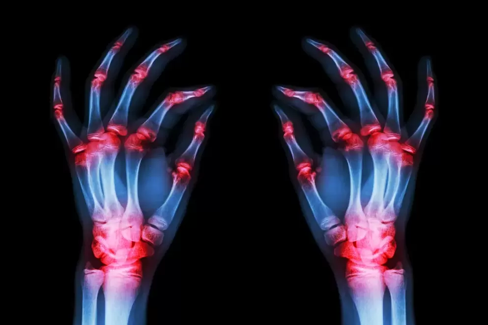
The main method for diagnosing joints is radiography. With arthrosis, changes in the joint, uneven joint surface and narrowing of the joint space can be observed.
Which joint is more likely to suffer from arthrosis?
The joints of the legs most susceptible to arthrosis are the hips, knees, shoulders, elbows and hands.
With arthrosis of the hip joint, a person may first feel some discomfort in the legs after running or walking. Over time, pain increases, limitations and stiffness in movement appear. With stage 3 disease, the patient protects his feet and tries, if possible, not to step on them.
Osteoarthritis of the knee joint manifests itself as pain in the knee joint after bending and straightening the leg. The most common cause of knee arthrosis is an injury suffered in the past. As a result of this injury, the gliding of the articular surface is disrupted and rapid wear occurs. In some cases, the joint may gradually lose its mobility.
Arthrosis of the ankle joint manifests itself in the form of swelling and pain in the ankle. The causes of arthrosis of the ankle joint can be: deformation, fracture of the ankle and talus, dislocation, flat feet, chronic injury of the ankle joint in athletes and ballerinas. In this way, they often suffer from foot arthrosis.
Arthrosis of the shoulder, elbow, and wrist joints most often appears as a result of injuries, bruises, dislocations, and intra-articular fractures. Arthrosis of the shoulder joint is characterized by pressing, aching, dull pain that radiates to the forearm and hand. Pain most often appears at night. With hand arthrosis, pain is accompanied by hand dysfunction.
Treatment of arthrosis
The main ways to treat arthrosis are drug treatment, the use of physiotherapy and surgical treatment.
Drug treatment
The use of drugs helps improve blood circulation in damaged joints, restore cartilage properties, and have analgesic and anti-inflammatory effects.
Nonsteroidal anti-inflammatory drugs
With arthrosis, joint swelling may appear, joints begin to ache and range of motion decreases. When taking anti-inflammatory drugs (NSAIDs), the pain is reduced, the inflammatory chain reaction is stopped and the cartilage recovery process is accelerated.
Medicines can be used in the form of tablets, rectal suppositories and powders. But remember that the drugs themselves are unacceptable; the selection and dosage of drugs for arthrosis is carried out by a rheumatologist.
Centrally acting pain relievers
Opioid drugs reduce the patient's pain threshold. Such drugs can be taken strictly according to the prescription and only under the supervision of a doctor!
Condoprotective drugs
Chondoprotective drugs are structural elements of cartilage itself, therefore they actively restore this tissue and prevent further destruction. Treatment is effective in the early stages of the disease. When the joint is completely destroyed, it is not possible to restore the original shape of the deformed bone or grow new cartilage.
However, at stage 1-2 arthrosis, chondroprotectors can bring significant relief to patients. Combined preparations, which include both glucosamine and chondroitin sulfate, provide better results than single component preparations.
Chondroitin sulfate and glucosamine sulfate
These drugs help slow the inflammatory response in the tissue, help reduce cartilage damage and reduce pain. Often, these 2 drugs are used together in treatment, because they have a cumulative effect, but they must be taken for 3-6 months.
Hyaluronic acid
Gives the viscosity and elasticity of synovial fluid. Helps the joints slide well. Therefore, doctors often prescribe injections of hyaluronic acid into the affected joints.
Physiotherapy treatment
Physiotherapy treatment may include:
- UHF therapy;
- magnetic therapy;
- low intensity laser irradiation;
- electrophoresis with drugs;
- phonophoresis (using ultrasound to introduce medicine into the site of inflammation).
Surgery
Surgical treatment is used to restore and improve joint mobility, as well as to remove part of the damaged cartilage or menisci.
Surgical treatment of arthrosis is used in extreme cases, when drug treatment does not produce results, when severe pain occurs, partial or complete immobility of the joint.
During arthroscopic surgery, it is possible to remove part of the cartilage affected by arthrosis, polish it to provide a smooth surface, remove debris and cartilage growth, and cut part of the damaged ligament.
Knee replacement
With this operation, the articular surface of the knee joint is replaced with metal or a combination of prostheses. The prepared plate mimics the surface of articular cartilage. Such prostheses are made of a special alloy; it does not cause a rejection reaction in the patient, does not oxidize, and does not injure the surrounding tissue.
Hip surgery for arthrosis
During this operation, partial removal of the cartilage and bone tissue of the pelvis and femur is performed. Usually, the head of the femur and the articular surface of the pelvic bone are removed and replaced with a metal or metal-ceramic prosthesis.
Diet for arthrosis
Excess body weight is the biggest enemy of your joints. Most patients with arthrosis of the hip and knee joints are overweight.
Therefore, for arthrosis, a properly selected diet is recommended. It is believed that jellied meat cooked in cartilage soup is beneficial for arthrosis. It contains a lot of collagen and cartilage structural components, which help restore cartilage tissue.
Dairy products, protein and calcium are beneficial. Animal protein is found in lean meat and fish, while vegetable protein is found in buckwheat porridge, beans and lentils. Boiled, stewed and steamed dishes are very healthy.
The best diet for joints is a diet with a slight excess of carbohydrates (preferably complex carbohydrates), fruits and vegetables, and adequate amounts of protein and calcium.
Prevention of arthrosis
Prevention of arthrosis, no matter how trivial, lies in a healthy lifestyle. If possible, try to be in the fresh air, move, walk barefoot on sand, green grass, and just dirt. This type of walking improves muscle function and improves blood circulation in the legs.
The use of physical therapy with various arm and leg swings, twists, and turns will provide proper support for your joints.
Patients often ask if alternative treatment for arthrosis is possible? Yes, folk remedies can help in the early stages of the disease, reduce pain and improve the general condition of the patient. But it does not replace following your doctor's instructions.